- Start a Study
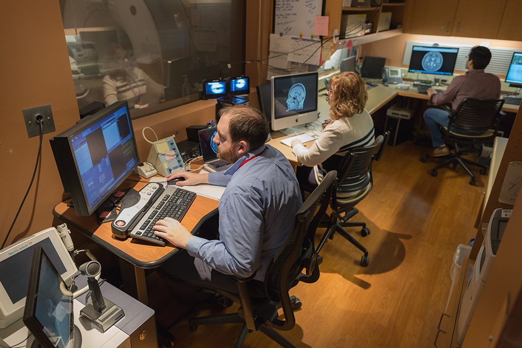
- Facilities
- Staff
- Affiliated Researchers
- FAQ
- Forms
- Resources for Researchers
- Contact Us
Getting Started:
- Complete the MRI Acquisition Strategy Request Form (at the bottom of this page) and direct any questions to Ginny Hayes, the Lane Neuroimaging MRI Lab Manager. The information on this form will help our trained staff assist you with the design of your experiment. The research question and experimental design are important aspects to obtaining useful data.
- Human Subjects Committee IRB approval is necessary before you can use the MRI at the Health Emotions Research Institute Lane Neuroimaging Lab. HSC/IRB approval can be a lengthy process, therefore it is important to submit your Protocol as soon as possible. Your proposed Consent Form should include all physiological measures you wish to collect, the ability to run two or three single session pilots to test both your paradigm and equipment, and information about “possible abnormal findings”.
Once you are approved:
- Once you have received approval from the IRB, complete the MRI Research Application (at the bottom of this page) and submit it along with your approved Protocol, approved Consent Form(s), and approved Funding/Grant information. You may upload these documents directly to the MRI Research Application below, or submit them to Ginny Hayes, the Lane Neuroimaging MRI Lab Manager. She will organize an MRI Users Committee meeting and notify you by letter of approval for MRI use. No scans or testing will be allowed without the MRI Users Committee approval.
- In addition to Study approval and documentation, it is also necessary for your research lab group members to obtain access to the Lane Neuroimaging MRI Suite. In order for this access to be given, each lab member must submit the following: the MRI Screening Form, verification of completion of the CITI training module(s) required by your IRB, and verification of completion of current HIPAA training. Depending on the medical information provided on the MRI Screening Form, the Surgical Procedure Information Form and/or a Surgical Implant Information Form may also be required.
- Finally, MRI safety is a serious matter. All persons involved in your research lab group who will be present during your scans will receive training to become Level 1 MR personnel. This means they must participate in the MRI Emergency training process and watch this MRI Safety Video and email Ginny Hayes, the Lane Neuroimaging MRI Lab Manager, when this has been done. In addition, your research lab members will be required to watch the MRI Safety Video as an annual Level 1 MRI safety refresher, probably at the same time the University requires annual HIPAA training be completed.Because it is not specific to the UW Clinics and Research program, a few points of clarification about the MRI Safety Video are needed:
- The examples of bad safety events are quite serious, and the one involving an infant might be a little unsettling. These examples are quite extreme, and very rare.
- The 5-gauss line is mentioned, which is a standard MRI concept. At HERI, the 5-Gauss line is not marked on the floor; it should be considered to be at the entry to the magnet room door.
- The video promotes changing all patients/subjects to hospital-provided clothing. We generally do not do this for research patients, but scrubs are provided if the MRI Techs believe a research subject’s clothing may be unsafe in the MRI.
- Research subjects are not given contrast agents for their MRI study.
- Once you have received your letter for approval from the MRI Users Committee and have completed the safety and access requirements for your research lab staff, you may email Ginny Hayes, the Lane Neuroimaging MRI Lab Manager, to learn about access to the MRI calendar, to discuss time slots for scanning, the equipment available for stimulus and data collection, and your MRI protocol. You may need to schedule time on the MR computer to work with Lane Neuroimaging Lab staff to set up your research protocol on the GE scanning computer, prior to your pilot scan session(s).
- The MRI Simulator is available for use if you choose to prepare your subjects with a simulation of an actual MRI scan. Please contact Ginny Hayes the Lane Neuroimaging MRI Lab Manager, for additional information on the Simulator or to schedule its use.
Research Subject Preparation and Documentation:
- As you screen and prepare potential research subjects for your MRI study, please go over the Day of Scan Information to educate them about their needed preparation for the scan.
- As you screen and prepare potential research subjects for your MRI study, complete the MRI Screening Form. Depending on the medical information provided on the MRI Screening Form, a Surgical Procedure Information Form and/or a Surgical Implant Information Form may also be required.
- On the day of your subject’s research scan, you must bring a copy of their MRI Screening Form (and MRI: Surgical Procedure Form and MRI: Surgical Implant Form, if required by their medical information) along with a copy of the signed consent form(s) to the MRI Control Room. All of this documentation will be reviewed and approved by the MRI technologist before you will be allowed to scan. The research subject may not enter the Magnet Room under any circumstances until given permission by the MRI technologist.
How to Schedule:
- For instructions on how to view the MRI Scanner and Simulator Calendars for scheduling, please contact Daniel Retzlaff.
- When you are able to view the MRI Calendar and have decided what time you would like to reserve for your study, please email your request to: Ginny Hayes, Tracy Patterson-Hegg, and Matt Gabriel. When you are able to view the Simulator Calendar, you may schedule your desired time directly on the Calendar.
Obtaining your data:
When you begin a new study at the Lane Neuroimaging Lab, visit IT to get your account set up and receive instructions on both data acquisition and the processing environment.
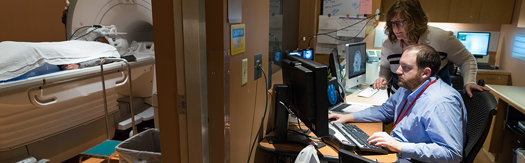
Facilities and Equipment
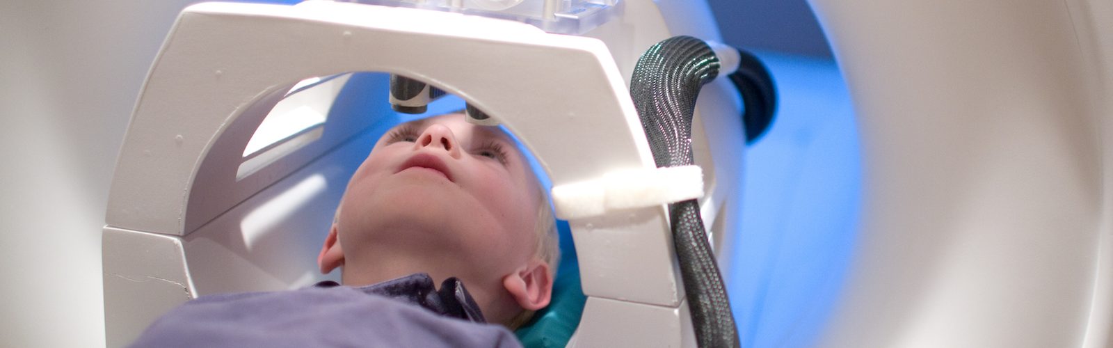
The Lane Neuroimaging Laboratory is located on the first floor of the Hedberg HealthEmotions Research Institute at the Wisconsin Psychiatric Institute and Clinics building. It houses a GE 3T scanner for structural and functional brain imaging, an MRI simulator room, an MRI preparation room for pre- and post-scan behavioral measures, image processing areas with extensive computing facilities, and a conference room and office space. All rooms at the Lane Neuroimaging Laboratory as well as at remote labs across the campus are networked together.
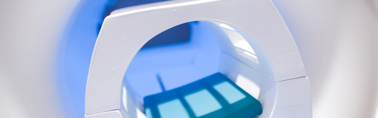
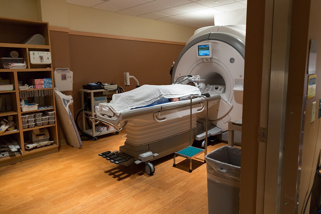
MRI Scanner
The Laboratory contains one of GE’s most advanced scanners, the 3.0 Tesla Discovery MR750. It is faster and provides greater image clarity than previous scanners. Researchers and clinicians can obtain images that were previously unimaginable.
GE’s EPIC software can be used to customize neuroimaging research. Stimulus Presentation can be delivered by using either an advanced fiber optic goggle system (by Avotec) or a Full HD (1920 x 1080p) DLP projector (by Resonance Technology). The projector can also be used together with an eye-tracker (by Avotec). This eye-tracking system yields four different analyses: percentage analysis, pictorial analysis, order of object vs. time, and pupil diameter vs. time.
MRI auditory stimulus is presented using a pneumatic headphone system (Avotec). All auditory stimuli are presented through a digital equalizer that is optimized for tone and clarity.
Stabilizing patients to minimize head movement is critical for MRI studies. The lab has two head stabilizing methods: vacuum pillow and foam inserts.
Collection of peripheral, physiological measures in the MRI are done using the Biopac MP160 system. The MP160 system provides high resolution, variable sample rates for analog and calculation channels, 16 analog inputs and two analog outputs, and 16 online calculation channels. The MP160 System provides high-speed acquisition (400 kHz aggregate), and Ethernet connectivity. Controlling the Biopac is the software, AcqKnowledge. This software is an interactive program that lets lab members instantly view, measure, analyze, and transform the incoming data.
The ability to collect both cardiac and respiration activity (sampling rate of 100 Hz and 25 Hz, respectively) is possible through the manufacturers standard software.
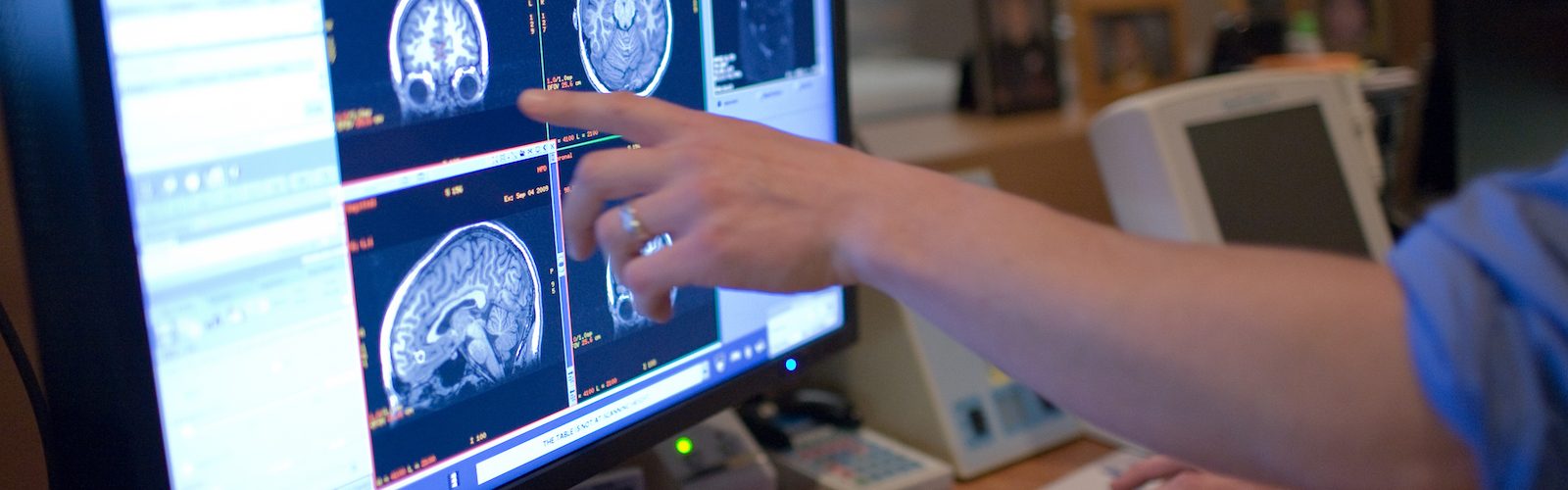
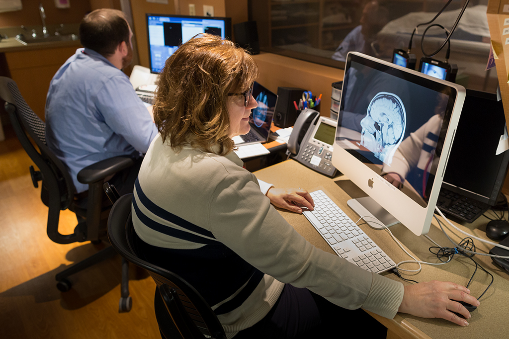
Stimulus presentation is controlled by E-Prime, Matlab, and NBS Presentation. Real-time monitoring of the image quality and subject motion is available via both FIRMM and AFNI. Morphometric measurements can use AFNI, FSL, Freesurfer, or SPM for coregistration. Both Freesurfer and in-house tools perform manual coregistration (BrainSqueezer) and distortion-based morphometry (DBM) measurements. Manual ROI drawing can use AFNI or an in-house tool (BrainMaker), automated ROI identification can be performed with Freesurfer. Talairach coordinates can be investigated using a large variety of standard atlases provided in AFNI and FSL. Diffusion Tensor Imaging (DTI) uses in-house software for analysis and display. Cortical flatmaps can be created using FreeSurfer.
Image Display is available through a variety of display programs, including AFNI, SPM, FSL, Freesurfer, Spamalize, LORETA, and BESA.
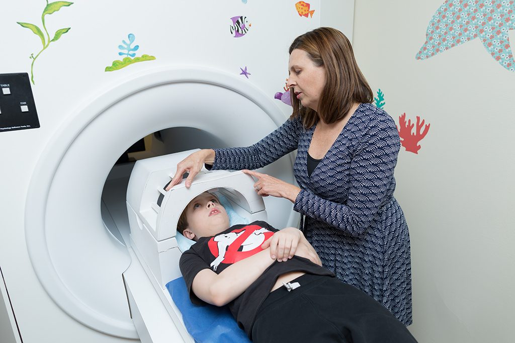
MRI Simulator
To acclimate research subjects to the MRI’s unique environment, the Lab provides an MRI Simulator. This MRI Simulator (non-magnetic) provides the experience and feel of an actual MRI scanner. The Simulator includes a moving table, lighting, fans, a stimulus projector system, and an auditory system with two control computers. The Simulator is used to introduce research subjects to the experimental procedures similar to what they will experience in the actual scanner, helping to ensure subject comfort and data quality.
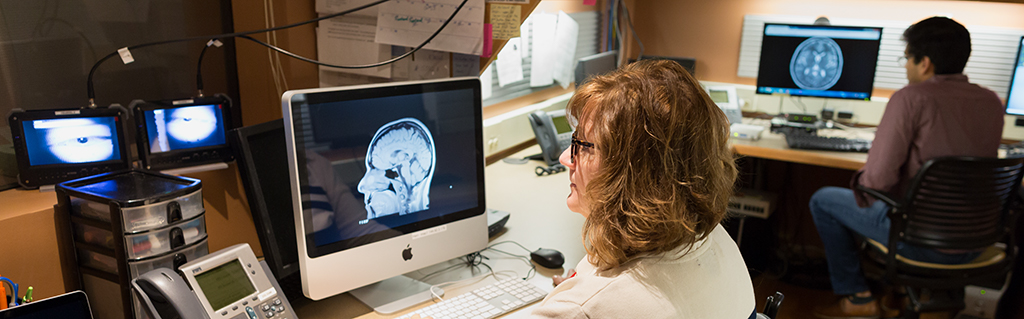
Computer Facilities
The HERI MRI computing network is split between two geographic locations: the local datacenter within the WISPIC building, and the datacenter at the Medical Foundation Centennial Building (MFCB). Both datacenters are secure and access controlled. All servers and storage equipment are on generator backed circuits, with UPS to provide power during cutovers.
The three processing servers are all Dell PowerEdge R840s. Each server has four Intel Xeon 26-core CPUs with multi-threading, providing 208 effective cores per system; the cores are clocked at 2.0GHz (3.7GHz turbo). In addition, each server has 512GB of RAM. Each server is equipped with 10GB networking for communication with the TrueNAS. Each server is running Ubuntu Linux and is routinely updated with the latest security patches.
The SAN is an iXSystems TrueNAS Z20, which utilizes the ZFS filesystem. The enclosure contains two physical nodes for redundancy, and each connect to the processing servers via 10GB networking. Equipped with dozens of HDDs in RAID 10, the system serves up ~300TB of raw storage. The system is equipped with multiple SSDs, providing both read and write caches for increased performance. Lab volumes are served up to the processing servers via the NFS protocol.
User workstations range from Apple iMacs to PC workstations, the latter usually containing a Linux distribution (typically Ubuntu). Authentication is handled through a redundant LDAP server, with user data being tightly access controlled across both servers and workstations.
Access to a wide variety of data analysis software is available throughout the lab. The following programming languages are actively used and supported: C/C++, Java, Perl, Python, Ruby, and R, among many others. Many industry-standard software suites are available, including Matlab, AFNI, and FreeSurfer; in most cases, any UNIX-compatible software can be installed on-demand to accommodate any use case. Multiple Standard office-related software packages are ubiquitous, including Microsoft Office and Adobe Photoshop/Illustrator/Acrobat.
Faculty and Staff

Ned H. Kalin, MD
Hedberg Professor, Chair, Department of Psychiatry
Director: HealthEmotions Research Institute
Director: Lane Neuroimaging Lab
608-262-7626

Rasmus M. Birn, PhD.
Assistant Professor, Physicist
Lane Neuroimaging Lab
Room 110b
608-265-5609
rbirn@wisc.edu
Ginny Hayes
Lab Manager
Room 1003
Lane Neuroimaging Lab
608-263-3278
vhayes@wisc.edu

Daniel Retzlaff
Network Administrator
Room 110
Lane Neuroimaging Lab
608-262-5944
deretzlaff@wisc.edu
Tracy Patterson-Hegg
MRI Technologist
Room 121
HERI MRI Control Room
608-232-3345
pattersonheg@wisc.edu
Matthew Gabriel
MRI Technologist
Room 121
HERI MRI Control Room
608-232-3345
mjgabriel2@wisc.edu
Affiliated Researchers
 Andy Alexander, Phd.
Faculty Co-Director, Brain Imaging Core
Associate Professor of Medical Physics & Psychiatry
Director of MR Physics Research, Waisman Laboratory for Brain Imaging and Behavior
Waisman Center
1500 Highland Avenue, Room T135
Madison, WI 53705
(608) 265-8233
aalexander@waisman.wisc.edu
 Rasmus M. Birn, PhD.
Assistant Professor, Psychiatry
HERI room 110b
608-265-5609
rbirn@wisc.edu
Ned H. Kalin, MD
Hedberg Professor, Chair, Psychiatry
Director: HealthEmotions Research Institute
Director: Lane Neuroimaging Lab
608-262-7626
 Mike Koenigs, PhD
Assistant Professor, Psychiatry
608-263-1679
mrkoenigs@wisc.edu
HERI room 1026A
 Jonathan Oler, PhD
Associate Scientist, Psychiatry
608-263-4021
oler@wisc.edu
HERI room 110
 David Plante, MD, PhD
Professor, Psychiatry and Sleep Medicine
dplante@wisc.edu
Wisconsin Sleep
 Brad Postle, PhD
Professor, Psychology and Psychiatry
608-262-4330
postle@wisc.edu
Brogden room 515
 Vivek Prabhakaran, MD
Assistant Professor, Neuroradiology and Psychiatry
608-265-5269
 Giulio Tononi, MD, PhD
Professor, Psychiatry
608-263-6063
gtononi@wisc.edu
HERI room 1334
Ryan Herringa, MD, PhD
Assistant Professor, Psychiatry
608-263-6068
herringa@wisc.edu

Melanie Boly, MD, PhD
Assistant Professor of Neurology
boly@neurology.wisc.edu

Doug Dean III
Assistant Professor of Neurology
deaniii@wisc.edu
 Stephanie Jones, PhD
Associate Scientist, Psychiatry
Assistant Director of Compliance and Outreach
Wisconsin Institute for Sleep and Consciousness
sgjones2@wisc.edu

Zachary Stowe, MD
Professor of Psychiatry
Director, Women’s Mental Health Program
zstowe@wisc.edu
FAQ
What hours is the MRI available to scan subjects?
The MRI is available for research scans Monday through Friday from 1:00 p.m. to 8:30 p.m. Generally, the MRI is also open two Saturdays a month, from 9:00 a.m. to 5:00 p.m. For further information, contact Ginny Hayes, the Lane Neuroimaging Lab Manager at (608) 265-4019 or by email at researchmri@psychiatry.wisc.edu.
The Simulator is available for use Monday through Friday from 8:00 a.m. to 8:00 p.m. Additional Saturday hours are available on Saturdays when the MRI is open for scanning. For further information, contact Ginny Hayes, the Lane Neuroimaging Lab Manager at (608) 265-4019 or by email at researchmri@psychiatry.wisc.edu.
What are the rates and rules for imaging?
The MRI Scanning fee is $600 per hour for all persons affiliated with the University of Wisconsin System and its Associates. There is a minimum one-hour charge for all scans. Additional time in the same scanning session will be billed in half hour increments.
A Radiology fee of $100 is charged for all over reads.
Once your study is approved, at your request, you will be scheduled for a specific time slot on the MRI calendar. Lab policy is that any time slot cancelled, within one week of the scheduled scan time or less, will be charged the entire session scan fee, unless another investigator requests to use that particular scan time.
When your study is approved, if you need time on the MRI scanner for pilot/testing to evaluate a technique for your study protocol, please contact Ginny Hayes, the Lane Neuroimaging Lab Manager at (608) 265-4019 or by email at researchmri@psychiatry.wisc.edu, to schedule this time.
Where is the Lane Neuroimaging Lab located?
We are on the main floor of the Hedberg HealthEmotions Building. The entrance is the modern glass addition on the far west end of the Wisconsin Psychiatric Institute & Clinics (WisPIC) building. Our physical address is 6001 Research Park Blvd, Madison, WI 53719.
Where do I park?
There is plenty of free parking in the lot surrounding the building.
Is there a bus stop nearby?
Yes, the West Transfer Point is ½ a block away on Tokay Blvd. and it offers all-day service. Please refer to the City of Madison Metro Schedule for more information.
Lane Neuroimaging Lab Forms
(links will open in a new tab)
- MRI Screening Form
To be completed by MRI subject or physician and sent via FAX or MAIL only. - Surgical Procedure Information Form
To be completed by MRI subject or physician and sent via FAX or MAIL only. - Surgical Implant Information Form
To be completed by MRI subject or physician and sent via FAX or MAIL only. - Day Of Scan Information
Information and instructions to help subjects prepare for MRI scan - COVID-19 Screening Form
Print & give to MRI Team on day-of-scan.
Resources for Researchers
Facility Statement
The Lane Neuroimaging Laboratory is located on the first floor of the Hedberg HealthEmotions Research Institute at the Wisconsin Psychiatric Institute and Clinics building. It houses a GE 3T scanner for structural and functional brain imaging, an MRI simulator room, an MRI preparation room for pre- and post-scan behavioral measures, image processing areas with extensive computing facilities, and a conference room and office space. All rooms at the Lane Neuroimaging Laboratory as well as at remote labs across the campus are networked together.
Rigor and Reproducibility at the Lane Neuroimaging Lab
The following steps are being taken at the Lane Neuroimaging Lab in order to assure scientific rigor and reproducibility.
Quality Assurance (QA)
• Quality Assurance (QA) scans of a phantom (gel filled sphere) are performed on a weekly basis across all of the radio-frequency (RF) coils used in research MRI scans.
• Quarterly maintenance of the MRI scanner is performed by the manufacturer, General Electric (GE).
• Annual quality testing and calibration of the MRI scanner are performed according to the American College of Radiology (ACR) guidelines.
Operation of the MRI scanner
• The MRI scanner for all research scans is operated by a trained MRI technologist
• Protocols for each MRI study are saved on the MRI scanner. This facilitates running the same set of data acquisition sequences, in the same order, for all subjects within each study.
• MRI technologists have a hard-copy of the protocol for each study with additional notes that is used as a guide during the MRI scanning session.
Training and Education
• New scanner technologists are trained by current/former scanner technologist, lab manager Ginny Hayes, and MRI physicist Rasmus Birn.
• Research teams are trained at the beginning of each study by the MRI techs and lab managers. This training includes how to correctly complete the MRI Safety Documents and how to comply and interpret the Day of Scan Form. These Forms are all created and reviewed by UW Radiology and the UW MRI Safety Committee. The techs also train Research Labs in how to complete MRI Emergency Procedures.
Documentation
• Information about every MRI scan is recorded in a log book. This information includes the date, exam number, study ID, PI name, the number of series in the scan, whether a Radiology overread was requested, and the number of backup discs used. This log book is stored in a locked cabinet in the MRI control room.
• MRI scanning parameters are stored in the header of each DICOM image
5 Most Recent Publications
- Heyn, S. A., Bailowitz, S., Russell, J. D., & Herringa, R. J. (2022). Sex‐based variations of prefrontal structure and longitudinal symptoms in pediatric posttraumatic stress disorder. Depression and Anxiety, 39(12), 902-912.
- George, G. C., Keding, T. J., Heyn, S. A., & Herringa, R. J. (2022). Longitudinal hippocampal circuit change differentiates persistence and remission of pediatric posttraumatic stress disorder. Depression and anxiety, 39(1), 8-18.
- Heyn SA, Keding TJ, Schmit C, Wolf RC, Herringa RJ. Neurobehavioral correlates of impaired emotion recognition in pediatric PTSD (2022). Development and Psychopathology, 34(3), 946-956.
- Moody JF, Aggarwal N, Dean DC 3rd, Tromp DPM, Kecskemeti SR, Oler JA, Kalin NH, Alexander AL. Longitudinal assessment of early-life white matter development with quantitative relaxometry in nonhuman primates. Neuroimage. 2022 May 1;251:118989. doi: 10.1016/j.neuroimage.2022.118989. Epub 2022 Feb 10. PMID: 35151851; PMCID: PMC8940652.
- Aggarwal N, Williams LE, Tromp DPM, Pine DS, Kalin NH. A dynamic relation between whole-brain white matter microstructural integrity and anxiety symptoms in preadolescent females with pathological anxiety. Transl Psychiatry. 2022 Feb 8;12(1):57. doi: 10.1038/s41398-022-01827-y. PMID: 35136030; PMCID: PMC8825837.
Top 10 Overall Users of the Core
- Ned Kalin, MD
nkalin@wisc.edu - Rasmus Birn, PhD
rbirn@wisc.edu - Josh Cisler, PhD
josh.cisler@austin.utexas.edu - Doug Dean, PhD
deaniii@wisc.edu - Ryan Herringa, MD, PhD
herringa@wisc.edu - Stephanie Jones, PhD
sgjones2@wisc.edu - Michael Koenigs, PhD
mrkoenigs@wisc.edu - David Plante, MD, PhD
dplante@wisc.edu - Brad Postle, PhD
postle@wisc.edu - Giulio Tononi, MD, PhD
gtononi@wisc.edu
Additional Resources
- Letter of Support Template for Grant Writers
- Line Drawing of our Core
- Rigor and Reproducibility Statement
Contact Us
Lane Neuroimaging Laboratory
HealthEmotions Research Institute
6001 Research Park Blvd.
Madison, WI 53719
608-265-4019
ResearchMRI@psychiatry.wisc.edu
If you have a question or if you are interested in participating in one of our numerous research studies, please contact us at 608-265-4019 or ResearchMRI@psychiatry.wisc.edu.
Forms
- MRI Screening Form
To be completed by MRI subject or physician and sent via FAX or MAIL only.
- Surgical Procedure Information Form
To be completed by MRI subject or physician and sent via FAX or MAIL only.
- Surgical Implant Information Form
To be completed by MRI subject or physician and sent via FAX or MAIL only.
- Day of Scan Information
Information and instructions to help subjects prepare for MRI scan
- COVID-19 Exposure Screening
Print and give to MRI Team on day of scan.